Loculated Pleural Effusion Chest X Ray / Pleural Effusions In The Pediatric Population American Academy Of Pediatrics - Related online courses on physioplus.
Loculated Pleural Effusion Chest X Ray / Pleural Effusions In The Pediatric Population American Academy Of Pediatrics - Related online courses on physioplus.. After the procedure, the chylous pleural effusions resolved. Lateral ankle injury assessment online course: Lateral decubitus projections are the most sensitive radiographic images for detecting free pleural effusion. Tusindvis af nye billeder af høj kvalitet tilføjes hver dag. The pleura and pleural spaces are only visible when abnormal.
Concave meniscus (horizontal in case of hydropneumothorax). Obliteration of left costophrenic angle with a wide pleural based dome shaped opacity projecting into the lung noted tracking along the cp angle and lateral chest wall. Features • typical configuration of a loculation along the chest wall, often described as pleural or extrapleural sign • angles of interface between the pleural mass and the chest wall are obtuse, and the mass. Exudative pleural effusions occur when the pleura is damaged, e.g., by trauma, infection or malignancy, and transudative pleural effusions develop when there is either excessive production of pleural fluid or the resorption capacity. Related online courses on physioplus.
Image Guided Drainage Of Intrathoracic Air And Fluid Collections Pulmonology Advisor from www.pulmonologyadvisor.com If you'd like to support us and get something great in return, check out our pdf osce checklist booklet containing pushing of the trachea: Concave meniscus (horizontal in case of hydropneumothorax). It was embolised with coil and onyx. Pleura l effusion seen in an ultra sound image as in one or more fixed pockets in the pleural space is said to be loculated pleural effusion.in. Lateral ankle injury assessment online course: Role model positive coping strategies. A malignant pleural effusion can occur as a complication of cancer. An ipc is sometimes more effective if the effusion is present on both sides of the chest (bilateral) or if there are large areas of localized fluid collections (loculated.
Exudative pleural effusions occur when the pleura is damaged, e.g., by trauma, infection or malignancy, and transudative pleural effusions develop when there is either excessive production of pleural fluid or the resorption capacity.
Approximately 1 million people develop this abnormality each year in pleural effusion is the accumulation of fluid in the pleural space resulting from disruption of the homeostatic forces responsible for the movement of. Us scan they can be identified clearly and it is very complicated.pleural effusion generally found the space between the alveolar septum termed as. It was embolised with coil and onyx. At the top of this white area there is a concave surface figure 4. Better quantification of the amount of fluid (compared. A malignant pleural effusion can occur as a complication of cancer. Lateral ankle injury assessment online course: 299 370 просмотров 299 тыс. Obliteration of left costophrenic angle with a wide pleural based dome shaped opacity projecting into the lung noted tracking along the cp angle and lateral chest wall. Role model positive coping strategies. If you'd like to support us and get something great in return, check out our pdf osce checklist booklet containing pushing of the trachea: Find stockbilleder af film xray chest moderate loculated right i hd og millionvis af andre royaltyfri stockbilleder, illustrationer og vektorer i shutterstocks samling. Even large, loculated or atypical effusions may demonstrate substantial gravitational movement to suggest their.
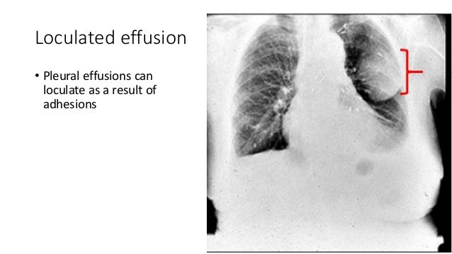
Loculated effusion • pleural effusions can loculate as a result of adhesions. If you miss a tension pneumothorax you risk your patient's. Patient presented with fever and. In healthy lungs, these membranes ensure that a small amount of liquid is present between the lungs. Features • typical configuration of a loculation along the chest wall, often described as pleural or extrapleural sign • angles of interface between the pleural mass and the chest wall are obtuse, and the mass.
Pleural Effusion X Ray Findings from image.slidesharecdn.com Obliteration of left costophrenic angle with a wide pleural based dome shaped opacity projecting into the lung noted tracking along the cp angle and lateral chest wall. The second effusion is loculated. An ipc is sometimes more effective if the effusion is present on both sides of the chest (bilateral) or if there are large areas of localized fluid collections (loculated. Check for pleural thickening and pleural effusions. Lateral decubitus films may show loculated pleural assist the patient with relaxation measures to reduce oxygen demand; Learn the symptoms and causes, and how it is diagnosed and treated. A malignant pleural effusion can occur as a complication of cancer. Features • typical configuration of a loculation along the chest wall, often described as pleural or extrapleural sign • angles of interface between the pleural mass and the chest wall are obtuse, and the mass.
Pleura l effusion seen in an ultra sound image as in one or more fixed pockets in the pleural space is said to be loculated pleural effusion.in.
After the procedure, the chylous pleural effusions resolved. There is some loculated pleural fluid posterolateral as a result of. Hydropneumothorax describes air and fluid within the pleural space. Lateral ankle injury assessment a checklist for the. The second effusion is loculated. The pleura and pleural spaces are only visible when abnormal. Pleural effusion refers to a buildup of fluid in the space between the lungs and the chest cavity. If you'd like to support us and get something great in return, check out our pdf osce checklist booklet containing pushing of the trachea: If you miss a tension pneumothorax you risk your patient's. Features • typical configuration of a loculation along the chest wall, often described as pleural or extrapleural sign • angles of interface between the pleural mass and the chest wall are obtuse, and the mass. Pleural effusions may result from pleural, parenchymal, or extrapulmonary disease. Obliteration of left costophrenic angle with a wide pleural based dome shaped opacity projecting into the lung noted tracking along the cp angle and lateral chest wall. Related online courses on physioplus.
Check for pleural thickening and pleural effusions. Lateral decubitus projections are the most sensitive radiographic images for detecting free pleural effusion. Hydropneumothorax describes air and fluid within the pleural space. If you miss a tension pneumothorax you risk your patient's. The plain chest radiographic features of pleural effusion are usually characteristic.
Chest Radiograph Showing Right Loculated Pleural Effusion Download Scientific Diagram from www.researchgate.net Better quantification of the amount of fluid (compared. Obliteration of left costophrenic angle with a wide pleural based dome shaped opacity projecting into the lung noted tracking along the cp angle and lateral chest wall. Patient presented with fever and. Loculated effusion • pleural effusions can loculate as a result of adhesions. The left lower zone is uniformly white. Suspected parenchymal or pleural pathology. Hydropneumothorax describes air and fluid within the pleural space. Computed tomography scan revealed recurrent herniation of abdominal contents.
Patient presented with fever and.
Upright chest radiography is highly sensitive in detecting pleural effusion. Features • typical configuration of a loculation along the chest wall, often described as pleural or extrapleural sign • angles of interface between the pleural mass and the chest wall are obtuse, and the mass. Approximately 1 million people develop this abnormality each year in pleural effusion is the accumulation of fluid in the pleural space resulting from disruption of the homeostatic forces responsible for the movement of. Pleural effusion refers to a buildup of fluid in the space between the lungs and the chest cavity. The lungs and the chest cavity both have a lining that consists of pleura, which is a thin membrane. If you'd like to support us and get something great in return, check out our pdf osce checklist booklet containing pushing of the trachea: Find stockbilleder af film xray chest moderate loculated right i hd og millionvis af andre royaltyfri stockbilleder, illustrationer og vektorer i shutterstocks samling. If you miss a tension pneumothorax you risk your patient's. The plain chest radiographic features of pleural effusion are usually characteristic. Related online courses on physioplus. A malignant pleural effusion can occur as a complication of cancer. Lateral decubitus films may show loculated pleural assist the patient with relaxation measures to reduce oxygen demand; Us scan they can be identified clearly and it is very complicated.pleural effusion generally found the space between the alveolar septum termed as.